
Optical coherence tomography (OCT) is a great, non-invasive imaging modality that many ODs are using in their practices. However, OCTs are often ordered only after abnormalities have been noted on fundoscopic examination, and thus there is a strong possibility that some of the early and subtle signs of geographic atrophy (GA) are being missed, says Julie Rodman, OD, MS, FAAO, chief of The Eye Care Institute in Broward, Florida and a professor at Nova Southeastern University College of Optometry.
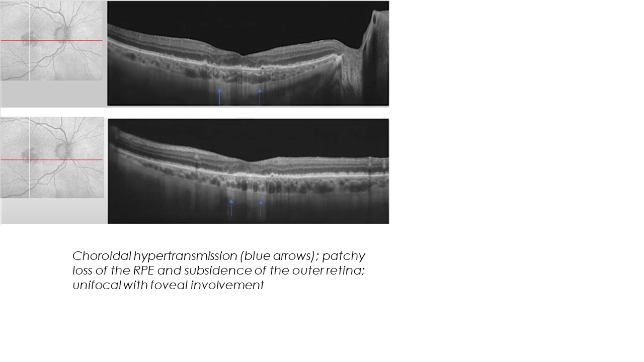
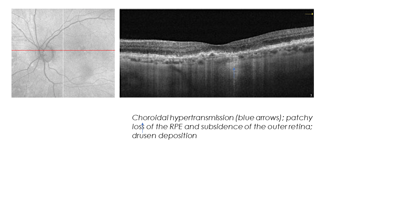
GA is the advanced form of dry age-related macular degeneration (AMD) and is defined by the presence of sharply demarcated atrophic lesions of the outer retina, retinal pigment epithelium and choriocapillaris.1,2
Fundus autofluorescence (FAF) and color fundus photography are other imaging modalities that are used in the diagnosis and management of geographic atrophy. However, OCT provides different but complementary information to these modalities, allowing the optometric community to do more to diagnose GA earlier. “If OCT was used more often as a wellness screener for patients with certain risk factors, we could do more to slow this progressive and irreversible disease,” she says.
“OCT allows precise visualization and characterization of the GA lesion. It helps identify the focality, location and extent of damage, which are not readily visible on color fundus photography.” — Dr. Julie Rodman
BE PROACTIVE
In her role as an educator and clinician, Dr. Rodman encourages practitioners to be more proactive in looking for signs of GA. “If patients have a family history of macular degeneration, they’ll get an OCT with me. Any patient who shows signs of drusen, I’ll want an OCT,” she says. The same is true with patients with pigmentary changes.
But even beyond the clinical signs, Dr. Rodman listens for patients to talk about particular symptoms, such as trouble seeing well in low levels of light. “If that is not related to cataracts, early GA might be a factor.” Dr. Rodman teaches in a medically oriented clinic, so it’s not uncommon to hear patients articulate complaints like this.
But these patients walk into most optometric offices on daily basis. So it’s important that optometrists are not only familiar with the symptomatology associated with macular degeneration but also the warning signs of the disease. It is very important optometrists become comfortable with grading macular degeneration. There are a lot of resources, she notes.
CE: There are courses on macular degeneration and GA at most meetings. In addition, attend CE courses that emphasize the use of OCT imaging.
OCT manufacturers: These companies hire representatives who are knowledgeable and able to consult. Many also collect case examples so that others can benefit from shared knowledge.
Industry: Companies like Iveric Bio are working to help more doctors understand the symptoms and clinical signs of GA with detailed information on their websites. These resources can also help eye care professionals become more comfortable knowing when to refer.
Iveric Bio has also created the Imaging Guide for Early Detection and Monitoring, an eight-page publication with photos and images that help explain what clinicians can look for.
 This level of emphasis will help create a “paradigm shift,” says Dr. Rodman, not unlike the shift that occurred in the past few years with more primary care optometrists becoming more proactive with children with progressive myopia.
THE R&D PIPELINE
The pharmaceutical industry is continuing to work on developments specifically for GA. Dr. Rodman notes that she often needs to remind students that the treatment for GA is not the same as that for neovascular age-related macular degeneration. Although both are intravitreal injections, neovascular AMD is managed with Anti-VEGF agents, while GA is being managed with complement factor inhibitors. These complement factor inhibitors do not reverse the disease, but they slow the progression, thus emphasizing the importance of early detection.
In April, Iveric Bio reported that in clinical trials in an interventional study, patients treated with a avacincaptad pegol suggested that a reduction of growth in GA resulted in an overall lower rate of vision loss. The data signaled a 56% risk reduction in the rate of persistent vision loss in GA patients.
In advance of treatment options coming onto the market, the optometric community can prepare, Dr. Rodman says. “This is in optometry’s wheelhouse. By the time that there’s foveal involvement, patients have been robbed of their central vision. The goal really should be to capture those patients who do not have foveal involvement and have not yet experienced central vision loss,” she says.
She encourages eye care professionals to take classes or read resources on how imaging can help detect early signs of the disease. “The hope is that we can catch patients while it’s not too late so that we can slow the progression and avoid that central vision loss and the psychological effects that this disease has on our patients.”
References:
- Fleckenstein M, Mitchell P, Freund KB, et al. The progression of geographic atrophy secondary to age-related macular degeneration. Ophthalmology. 2018;125(3):369-390.
- Sadda SR, Chakravarthy U, Birch DG, Staurenghi G, Henry EC, Brittain C. Clinical endpoints for the study of geographic atrophy secondary to age-related macular degeneration. Retina. 2016;36(10):1806-1822.
Read other stories about how ODs are detecting and talking with patients about GA here.
This content is independent editorial sponsored by Astellas. Astellas had no input in the development of this content. Astellas, formerly Iveric Bio.

